Skip to content

A Lightweight Explainable AI Framework for Early Brain Tumor Detection Using EEG Functional Connectivity

Aug 2026 · Science Engineering and Technology · 0 citations · 44 references

TL;DR

The findings support the feasibility of lightweight graph-based EEG analysis as an auxiliary screening and decision-support approach and at the same time, MRI and CT remain necessary for clinical confirmation and anatomical characterization.
Abstract

Early identification of brain tumors is an important technique for timely clinical assessment, but magnetic resonance imaging (MRI) and computed tomography (CT) remain the primary modalities for anatomical confirmation and tumor localization. Electroencephalography (EEG) provides a non-invasive and comparatively accessible complementary modality that can capture functional alterations in neural activity. This study presents Graph EEG Net-Lite, a lightweight and explainable graph neural network for auxiliary brain tumor screening using resting-state EEG functional connectivity. EEG recordings from 50 subjects, comprising 25 participants with radiologically confirmed brain tumors and 25 neurologically healthy controls, were represented as functional connectivity graphs using 19 EEG channels. Signals were filtered using a zero-phase fourth-order Butterworth band-pass filter from 0.5 to 45 Hz and a 50 Hz notch filter, segmented into 4-s non-overlapping epochs, and represented using relative spectral power and phase-locking value (PLV)-based connectivity. Graph EEG Net-Lite employs two Chebyshev graph convolution blocks with 64- and 32-dimensional hidden representations, followed by global mean pooling and a shallow classifier. Under subject-independent evaluation, the model achieved 96.2% accuracy, 96.8% sensitivity, 95.5% specificity, 95.9% F1-score, and an AUC of 0.987. The model used 74.8% fewer parameters and required 61.3% less training time than the standard GNN baseline. GNNExplainer identified informative contributions from frontal, temporal, parietal, and occipital electrode regions. The findings support the feasibility of lightweight graph-based EEG analysis as an auxiliary screening and decision-support approach. At the same time, MRI and CT remain necessary for clinical confirmation and anatomical characterization.

Read PDF

Similar papers

Sep 2026

Mixture-of-Experts Electroencephalography-Functional Magnetic Resonance Imaging Fusion for Interpretable Computer-Assisted Brain Disorder Diagnosis.

Electroencephalography (EEG) and functional magnetic resonance imaging (fMRI) provide complementary information about brain function, and have shown significant promise in detecting functional abnormalities in various brain disorders. However, the distinct signal characteristics and disparate representational spaces of...

Rui-Zhe Zhong, Hai-Sheng Li, Qing-Chuan Zhang · 0 citations
#graph neural networks Open access Oct 2026

Early disease detection using multiscale EEG graph fusion and bioinspired deep ensemble learning

Early detection of Alzheimer’s disease (AD) is essential for timely clinical intervention, yet remains challenging due to subtle neural changes in its initial stages. This study proposes a non-invasive EEG-based framework that integrates multiscale signal processing, dynamic brain connectivity analysis, nonlinear fea...

G. Vinuja, S. Arunachalam, N. Ashok · 0 citations
Open access Sep 2026

BRAINet: a brain-region-aware interaction network for EEG-based diagnosis of disorders of consciousness

Objective. Reliable assessment and stratification of disorders of consciousness (DOC) is essential for patient care and clinical treatment planning. Electroencephalography (EEG) provides a non-invasive approach to measure neural activity and has shown promise in DOC assessment. However, most existing EEG-based approach...

Hao-Xiang Chen, Sha Zhao, Jie Yu et al. · 0 citations
Open access Oct 2026

An explainable EEG connectome framework for frequency-resolved analysis of functional connectivity alterations in Parkinson’s disease

Parkinson’s disease (PD) is a neurodegenerative disorder characterized by the degeneration of dopaminergic neurons, leading to a variety of motor and non-motor symptoms. Analyzing brain functional connectivity is crucial for understanding PD, with electroencephalography (EEG) serving as a valuable, non-invasive too...

Donato Romano, M. Magarelli, P. Novielli et al. · 0 citations
Conference Aug 2026

Detection of Schizophrenia Using Electroencephalography Signals with a Hybrid EEGNet and Transformer Model

The diagnosis of schizophrenia has historically been based on clinical opinion, a method which has been observed to lend itself to late diagnosis or non-standard outcomes. EEG offers an affordable, non-invasive window into brain dynamics that may support the development of objective biomarkers for psychiatric disorders...

Yeddula Sai Sreesha, P. N. Reddy, Y. Ramadevi et al. · 0 citations
Sep 2026

Multidomain Electroencephalography Biomarker Fusion for Machine Learning-Based Dementia Classification.

Electroencephalography (EEG) has been investigated as a noninvasive approach for characterizing brain activity in neurodegenerative conditions. This study evaluated whether multidomain EEG biomarkers could distinguish Alzheimer's disease (AD), frontotemporal dementia (FTD), and healthy controls (HC). The publicly avail...

Jobin Thomas, Sweet Subhashree, S. Mohanty et al. · 0 citations

Related blog posts

Microsoft Research Blog Jul 13, 2026

Verifying Rust cryptography in SymCrypt, from standards to code

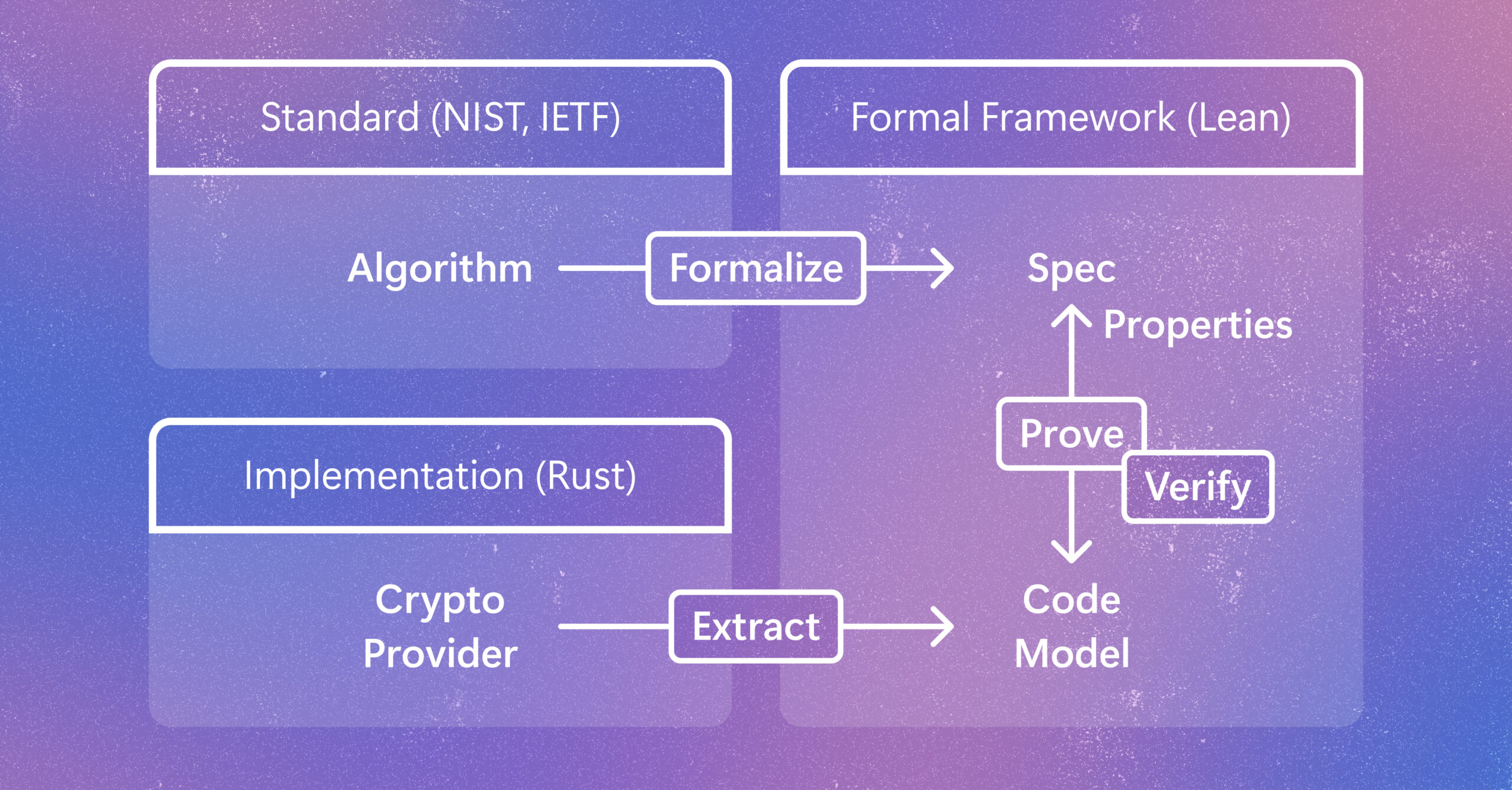
Cryptographic code supports vital protections in modern computing systems. Learn how a new method helps verify code as developers write it while preserving speed and adaptability as it gets implemented and evolves. The post Verifying Rust cryptography in SymCrypt, from standards to code appeared first on Microsoft Research.

We use cookies to run the site and, with your consent, for analytics and to show ads. See our Cookie Policy.