Aug 2026· Fluids and Barriers of the CNS· 0 citations
TL;DR
In vivo evidence is provided for choroid plexus dysfunction in a maternally inherited disorder with convergent evidence supporting a mitochondrial aetiology, associated with reduced CSF production and impaired blood–CSF barrier transport, substantially altering biomarker profiles in the absence of cortical atrophy or blood–brain barrier disruption.
Abstract
Choroid plexus dysfunction has been proposed to reduce CSF production and impair brain solute clearance, but the in vivo consequences of sustained clearance failure for biomarker profiles and disease mechanisms remain poorly defined. We examined Skogholt’s disease, a rare, maternally inherited neurodegenerative syndrome as a natural human model to test whether primary choroid plexus failure can reshape CSF dynamics and biomarker profiles independently of overt neurodegeneration.
Multimodal assessment of 40 individuals (mean age 57.8 years; 16 affected individuals and 24 controls) was used to evaluate clearance dynamics, diffusion properties, and CSF–plasma biomarker relationships, including model-free analysis of dynamic contrast-enhanced MRI washout of the choroid plexus.
We observed convergent evidence for a choroid plexus-centred clearance defect. Affected individuals had markedly reduced choroid plexus volume (Cohen’s |d| = 2.64) and prolonged contrast washout (time-above-half-peak |d| = 4.9) without cerebral parenchymal blood–brain barrier disruption. Diffusion MRI showed widespread reductions in mean diffusivity (|d| ≈ 1.0 in white matter; 0.85–0.89 in cortex), opposite to changes typical of neurodegeneration. CSF tau, amyloid β species, β-trace, neurofilament light and glial fibrillary acidic protein were elevated up to 12-fold, whereas plasma levels were normal or reduced, indicating marked CSF–plasma dissociation. Directional trace-metal shifts suggested impaired ATP-dependent transport. Mitochondrial genome sequencing identified a rare homoplasmic
MT-RNR2
(m.1681G > A) variant that segregates with disease and is absent as a homoplasmic variant in public mitochondrial reference datasets. The variant localises to a highly conserved structural element of the mitochondrial 16S ribosomal RNA, supporting its candidacy as a pathogenic variant pending functional validation.
These findings provide in vivo evidence for choroid plexus dysfunction in a maternally inherited disorder with convergent evidence supporting a mitochondrial aetiology, associated with reduced CSF production and impaired blood–CSF barrier transport, substantially altering biomarker profiles in the absence of cortical atrophy or blood–brain barrier disruption. Elevated CSF but normal or low plasma neurodegeneration markers most parsimoniously reflect impaired CSF-to-blood clearance, highlighting a clearance-sensitive dimension of biomarker biology. Dynamic contrast-enhanced MRI choroid plexus washout and CSF–plasma coupling emerge as complementary in vivo markers of clearance capacity. This syndrome may represent a prototype of disorders defined by primary CSF clearance failure rather than neuronal degeneration.

Highlights What are the main findings? Larger choroid plexus volume was associated in several studies with poorer cognition in MS, particularly processing speed and visuospatial memory. Longitudinal evidence did not show consistent predictive value for choroid plexus volume; one study suggested prognostic value of the CP T1/T2 ratio. What are the implications of the main findings? Choroid plexus volume and microstructural measures are promising MRI research markers, but not yet established clinical biomarkers. Harmonized acquisition and segmentation and adequately powered longitudinal, multidomain studies are needed. Abstract Background/Objectives: Multiple sclerosis (MS) is frequently accompanied by cognitive impairment, yet the neurobiological mechanisms underlying cognitive heterogeneity remain incompletely understood. The choroid plexus (CP), a blood–CSF barrier structure involved in cerebrospinal fluid production and neuroimmune signaling, has recently emerged as a potential MRI marker of inflammatory and neurodegenerative activity in MS. This scoping review mapped evidence on associations between CP features and cognitive functions in adults with MS. Methods: A broad search of PubMed, Scopus, Web of Science Core Collection, and the Cochrane Library identified 1163 records; after deduplication, screening, and full-text assessment, seven studies were included. Results: CP volume or normalized CP volume was assessed in all seven studies, and one study additionally examined the CP T1/T2 ratio. The Symbol Digit Modalities Test was used in six studies, while five applied multidomain cognitive assessment. Larger CP volume was associated in several studies with poorer baseline information-processing speed, visuospatial memory, or multidomain cognition, but longitudinal findings did not show consistent predictive value for CP volume. One study reported that a higher CP T1/T2 ratio predicted faster visuospatial-memory decline. Conclusions: CP-related measures may reflect broader neuroinflammatory and neurodegenerative processes relevant to cognition in MS, but the evidence remains limited and methodologically heterogeneous. Standardized acquisition and segmentation, harmonized cognitive assessment, and adequately powered longitudinal studies are needed to establish their independent and predictive value.
Weronika Galus, Patrycja Romaniszyn-Kania, Aleksandra Urantówka et al.· Brain Science· 0 citations
Parkinson's disease (PD) is a progressive neurodegenerative disorder commonly accompanied by cognitive decline, yet the mechanisms linking disrupted brain homeostasis to progressive cognitive impairment remain unclear. Emerging evidence suggests that the choroid plexus (ChP) and the locus coeruleus (LC) are involved in cerebrospinal fluid dynamics and norepinephrine regulation, respectively, but their longitudinal alterations and interrelationships in PD have not been systematically examined. We conducted a two-year longitudinal study including 90 PD patients and 51 healthy controls (HCs) undergoing multimodal MRI. ChP volume (ChP-V) was derived from T1-weighted structural imaging, ChP blood flow (ChP-BF) was assessed using pseudo-continuous arterial spin labeling, and LC integrity was ascertained with the contrast-to-noise ratio of the LC (LC-CNR) in neuromelanin-sensitive MRI. Group differences, longitudinal alterations, and associations with neuropsychological performance were examined. Over two years, PD patients showed progressive increases in ChP-V (F = 12.45, p < 0.001), reductions in ChP-BF (F = 18.98, p < 0.001), and declines in LC-CNR (F = 16.80, p < 0.001). Baseline LC-CNR was already reduced in PD compared with HCs (t = 3.023, p = 0.003). The longitudinal changes were more pronounced in male patients. ChP-BF was positively correlated with LC-CNR at baseline (r = 0.293, p = 0.006). Moreover, reductions in ChP-BF and LC-CNR were associated with worsening cognitive performance. While LC dysfunction was evident early in the disease course, progressive ChP alterations, particularly in ChP perfusion, provided additional information on longitudinal disease progression, supporting their combined value and highlighting the importance of gender-specific longitudinal monitoring.
H. Li, J. Jia, J. Wang et al.· medRxiv· 0 citations
BACKGROUND
Growing evidence supports choroid plexus (CP) involvement in MS pathophysiology, but phase-specific associations according to recent inflammatory activity and longitudinal dynamics remain unclear.
OBJECTIVE
To characterize CP alterations in MS using volumetric and quantitative magnetic resonance imaging (MRI) and explore associations with neuroinflammation and neurodegeneration.
METHODS
We analyzed cross-sectional and 2-year longitudinal data from 101 patients with active relapsing MS (RMS), 83 with inactive progressive MS (PMS), and 100 controls, with MS groups stratified by inflammatory activity within the prior year. CP volume, quantitative T1, and magnetization transfer saturation (MTsat) were assessed alongside imaging, serum, and clinical measures, with validation in a second cohort (62 active/357 inactive MS).
RESULTS
CP volume was increased in both active RMS and inactive PMS and was associated with lower total and cortical brain volumes. Associations with brain microstructural damage were more pronounced in periventricular regions, compatible with a surface-in pattern. Similar associations were observed in controls, suggesting CP volume may reflect processes beyond MS-specific pathology, including physiological factors. Longitudinally, CP volume increased exclusively in active RMS, with faster expansion associated with thalamic atrophy. CP MTsat was associated with cortical and paramagnetic rim lesions in inactive PMS.
CONCLUSION
CP enlargement is consistent across MS phases. Longitudinal expansion occurs in active inflammatory disease and is associated with neurodegenerative changes.
M. Mastantuono, A. Cagol, M. Ocampo-Pineda et al.· Multiple Sclerosis· 0 citations
Hydrocephalus is common with a mixed phenotype and aetiology. Its treatment is predominantly surgical, with temporary or permanent cerebrospinal fluid (CSF) diversion, which does not address the underlying disease mechanism. The choroid plexus (ChP), a secretory epithelium found in all four ventricles of the brain, produces most CSF within the central nervous system and has been investigated extensively over the past century to understand its function and potential role as a therapeutic target. Past attempts at either medically or surgically controlling its rate of secretion have not significantly altered our approach to the treatment of hydrocephalus, with CSF diversion remaining the main intervention. Rodent models of post-haemorrhagic hydrocephalus (PHH) have advanced our understanding of choroid plexus function in health and disease. Pre-clinical experiments have demonstrated a hypersecretory response in the ChP that may contribute to PHH. Targeting the choroid plexus directly may therefore present a novel therapeutic method. These findings have led to a significant increase in pre-clinical studies exploring this hypersecretory response and how to modulate it. Translating these promising results to clinical practice will rely on the development of large animal hydrocephalus models, which thus far has been limited. This literature review discusses the recent advances in targeting the ChP as a treatment for hydrocephalus, both surgically and non-surgically, and the current barriers to further advancement of this approach.
Olivier Sluijters, J. Pooley, W. Singleton· Fluids and Barriers of the C...· 0 citations
The choroid plexus (CP) plays a crucial role in cerebrospinal fluid secretion and the maintenance of brain homeostasis. Its structure and function have been implicated in the pathogenesis of dementia; however, the longitudinal associations of CP with dementia and structural brain biomarkers remain unclear. This prospective cohort study utilized data from the UK Biobank, including 45,306 participants (mean age, 64 years; 47.2% men) who underwent 3.0 T multiparametric brain MRI scans. CP volume and signal intensity were quantified by FreeSurfer software. Measures of grey or white matter macrostructures or microstructures were derived from structural or diffusion MRI. Dementia outcomes were identified using linkage of hospital admission records or death register. Tests for global and domain-specific cognitive functions were administrated at baseline and follow up. Data were analyzed using Cox proportional hazards, Mendelian randomization, linear mixed-effects models, and mediation models. Advancing age was correlated with increased CP volume (R = 0.49, P < 0.001) and decreased CP signal intensity (R = -0.44, P < 0.001). Meanwhile, greater CP volume was associated with an increased risk of all-cause dementia (hazard ratio, 1.61; 95% confidence interval, 1.32-1.97), while higher CP intensity was correlated with a reduced dementia risk (0.44; 0.36-0.55). Reduced volumes of the hippocampus, amygdala, and nucleus accumbens, increased white matter hyperintensity volume, mean diffusivity, and isotropic compartment volume fraction, and decreased intracellular volume fraction significantly mediated up to 42.9% of these associations. This study provides evidence supporting a causal relationship between CP morphological parameters and dementia risk that is partly mediated by specific brain phenotypes, and further suggests that the CP parameters may be valuable biomarkers for structural brain aging and dementia.
Alzheimer’s disease (AD) is usually regarded as a neurodegenerative disorder defined by amyloid-β (Aβ) deposition and abnormal tau accumulation. Increasing evidence suggests that reduced clearance of metabolic waste and pathological proteins from the brain also contributes to disease onset and progression. Previous studies have often considered choroid plexus (CP) function, glymphatic exchange, and meningeal lymphatic drainage as separate clearance processes. A continuous framework linking these structures and functions is still lacking. This review integrates recent clinical and experimental evidence and proposes the brain clearance continuum as an interpretative framework. It describes three interlinked functional interfaces: the upstream choroid plexus–cerebrospinal fluid (CP–CSF) inflow interface, the midstream parenchymal perivascular exchange interface, and the downstream meningeal lymphatic outflow interface. Under physiological conditions, these interfaces support CSF movement, parenchymal solute exchange, and the outward removal of metabolic waste. In AD, disrupted CSF homeostasis, impaired perivascular exchange, and obstructed meningeal lymphatic outflow may interact, leading to serial failure of the brain clearance continuum. This process is closely associated with Aβ/tau accumulation, vascular dysfunction, neuroinflammation, and cognitive decline. We also summarise potential therapeutic strategies directed at different clearance interfaces, whilst emphasising that most evidence remains preclinical or exploratory. The brain clearance continuum provides a systematic framework for understanding clearance failure in AD. It may also offer a theoretical basis for future mechanistic studies and therapeutic development that are stratified by clearance interface and disease stage.
Qi Yao, Xiaodong Han, Tong Cui et al.· Journal of Neurology· 0 citations
Related blog posts
MIT News · Artificial Intelligence· news.mit.eduAug 27, 2026
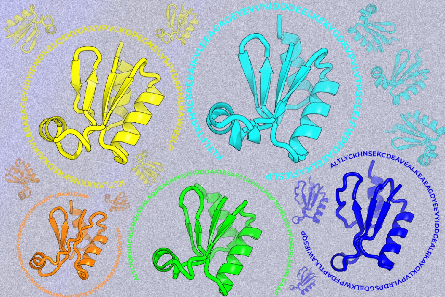
A new machine-learning framework aims to improve the success rate of computational protein design while moving away from results that reproduce sequences found in nature.