This is the first study reporting the use of phage/bacterium system and aggregation induced emission luminogen for detection of antibiotic residues in food sample.
Abstract
Background
For the previous rapid screening methods, antibody, molecularly imprinted polymer, aptamer and receptor are the usually used recognition reagents. It is well known that there are many antibiotic receptors on bacterial surface, so in theory the whole bacterium can be used as recognition reagent for screening of the specific antibiotic. However, such a method has not been reported so far. The aim of the present study is to develop a method for rapid screening of beta-lactam drugs in milk with Escherichia coli as the recognition reagent and with aggregation induced emission luminogen as the signal source.
Results
In this study, T4 phage was coupled with Fe3O4 to synthesize a type of magnetic complex that could specifically capture E. coli. Cephalexin was coupled with an aggregation induced emission luminogen (tetraphenylethylene) to synthesize a fluorescent tracer that could specifically bind with the penicillin binding proteins on E. coli surface. The magnetic complex, the E. coli solution and the tracer were mixed with beta-lactam drugs to perform competition. The fluorescent signal from the tracer molecules aggregated on bacterial surface was negatively correlated with the drug concentration. This method could be used for multi-screening of 28 beta-lactam drugs. The operation was simple and rapid, and the sensitivities for these drugs (limits of detection of 0.73-95.19 pg/mL) were improved for 16-959 folds in comparison with fluorescein based fluorescent tracer. The detection results for the real milk samples were consistent with a LC-MS/MS method.
Significance
This is the first study reporting the use of phage/bacterium system and aggregation induced emission luminogen for detection of antibiotic residues in food sample. Under the guidance of this study, more similar or advanced methods based on bacteria and novel signal sources for detection of antibiotics should be reported in the future.
Reliable detection and fast inhibition of Escherichia coli (E. coli) are critical for public health and food safety, so the detection-inhibition-integrated strategies become critical. Herein, a mannose-engineered 3,5-dicarboxyphenylboronic acid/curcumin dual-ligand Eu-metal-organic framework (Eu-MOF) was designed as dcMOF@man. Mannose realized the specific identification to E. coli; Eu-MOF exhibited dual-emission at 350 and 633 nm for robust ratiometric fluorescence sensing to E. coli as boronic acid-surface glycan interaction enhanced the emission at 350 nm, while mannose-FimH-mediated interaction attenuates the Eu3+ emission at 633 nm. The intensity ratio of I633/I350 exhibits a good linear relationship to E. coli at the concentration range of 10-107 CFU/mL with a low detection limit of 5 CFU/mL. Concurrently, the dcMOF@man exhibited peroxidase-like activity, enabling E. coli-dependent colorimetric detection based on tetramethylbenzidine oxidation, with the linear range of 10-107 CFU/mL and detection limit of 9 CFU/mL, which was also quantified via smartphone-based RGB analysis for on-site detection. The intrinsic antibacterial activity of curcumin afforded the effective inhibition of E. coli. Moreover, curcumin-mediated antibacterial activity synergizes with mannose targeting, resulting in nearly 100% antibacterial efficiency. Thus, dcMOF@man integrates targeting identification, ratiometric fluorescence sensing, colorimetric detection, and antibacterial functionality within single composite, as a generalizable strategy for on-site bacterial detection and inhibition.
Weiqi Wu, Zhenyu Wu, Sihang Yuan et al.· Talanta: The International J...· 0 citations
Antimicrobial resistance has emerged as a significant threat to global public health, necessitating accurate and rapid detection methods. To address this need, a colorimetric nanobiosensor was developed that exploits antibiotic-induced metabolic changes in bacteria, detected via the redox-dependent activity of a peroxidase-mimicking nanozyme (γ-Fe₂O₃@Prussian blue). This study aimed to differentiate resistant from susceptible E. coli strains and determine the minimum inhibitory concentration for susceptible ones using this novel principle. The experiments were conducted on two susceptible and two resistant strains of E. coli along with antibiotics, including ampicillin, cefazolin, ceftriaxone, and kanamycin. The γ-Fe2O3@PB NPs were characterized using UV-vis spectroscopy, dynamic light scattering (DLS), X-ray diffraction (XRD), transmission electron microscopy, and Fourier-transform infrared spectroscopy (FTIR). The synthesis of cubic crystalline nanoparticles with average crystallite size and hydrodynamic size of 31 nm and 219 nm, respectively, was confirmed. Upon adding H₂O₂ and the chromogenic substrate TMB to the bacterial culture supernatant, the intensity of the blue color (measured at 652 nm) in sensitive strains correlated with antibiotic concentrations. Compared to sub-MIC concentrations, a significant and sharp increase in absorbance at 652 nm was observed at the MIC, and similarly high absorbance levels were maintained at higher concentrations. By comparing the absorbance levels below and at the MIC, the MIC range can be determined. In contrast, for resistant strains, the intensity of the produced color remained nearly constant across different antibiotic concentrations. This innovative, label-free approach offers a simple and reliable method for antibiotic susceptibility testing. It achieves results in approximately 4 hours, with a detection limit (initial bacterial count) of 1.67×10⁷ CFU/mL, without requiring expensive reagents or equipment.
Staphylococcus aureus (S. aureus) is a common foodborne pathogen that can cause the severe contamination of dairy products. Therefore, there is an urgent need for rapid detection methods. In this study, a paper-based biosensor integrating a dual-recognition strategy using aptamers and antibodies was developed for the sensitive, rapid, and on-site detection of S. aureus in complex milk matrices. The biosensor combines a milk matrix-adapted aptamer with polyclonal antibodies (pAbs) and utilizes colloidal gold nanoparticles (AuNPs) as visual signal reporters. Using a SELEX process tailored to the milk matrix, the high-affinity aptamer SA2-1 was selected to specifically bind S. aureus, while pAbs enabled multi-epitope capture on the paper substrate. The aptamer–AuNP conjugates generated visual signals, achieving a detection limit of 102 CFU/mL within 15 min without an instrument. This dual-recognition strategy synergistically enhances both sensitivity and specificity, offering a cost-effective solution for dairy safety monitoring.
Wei-Chen Hou, Xiangyang Li, Jie Li et al.· Biosensors· 1 citation
Rapid, selective detection of bacterial pathogens remains a central challenge. Here we report a label-free electrochemical biosensing approach that leverages protein-protein interaction (PPI)-derived peptides as recognition elements for rapid detection of Listeria monocytogenes (LM). The sensor design is inspired by the interaction between the LM virulence factor Internalin A (InlA) and the human host receptor E-cadherin (E-Cad1). Peptides derived from the InlA-binding domain of E-Cad1 were engineered as molecular recognition elements, with the E-Cad1(15-24) peptide displaying micromolar affinity and selective binding towards LM. Immobilization of these peptides on gold electrodes enabled bacterial detection by electrochemical impedance spectroscopy within 10 minutes, without labels or external signal amplification. A low peptide surface density was associated with enhanced binding-site accessibility and may facilitate multivalent interactions between the bacterial surface and the immobilized peptides. The platform produced a detectable response at experimentally tested concentrations as low as 1 CFU mL ¹ and exhibited excellent selectivity under the conditions examined. This work introduces a chemically programmable, PPI-inspired biosensing paradigm that uses a reductionist approach and could potentially be extended to other pathogen targets.
Revital Krispin, Hen Okshtein, Yawen Song et al.· bioRxiv· 0 citations
ABSTRACT The escalating antimicrobial resistance crisis demands innovative strategies for antibiotic discovery. Conventional approaches for identifying antibiotic-producing microorganisms from environmental samples are often laborious and low-throughput, requiring prior isolation and purification of individual strains. Here, we developed a high-throughput screening platform integrating a microbial whole-cell biosensor into a double-layer plate assay, enabling rapid identification of bacteria producing cell wall-targeting antibiotics from environmental samples. The biosensor is based on the PghKR two-component system from the gram-negative bacterium Shewanella oneidensis MR-1. Upon exposure to cell wall-targeting antibiotics, PghKR activates the promoter of blaA, driving expression of the luxCDABE reporter and generating luminescence. A highly sensitive biosensor was engineered through the synergistic deletion of blaA and ampG, which greatly improved its responsiveness. The method was then applied to screen soil samples. From the primary screen, 103 colonies producing distinct luminescent signals were identified. Of these, 36 isolates consistently activated the biosensor in a confirmation assay, and 5 exhibited antibacterial activity against a multidrug-resistant indicator strain. This integrated approach combines microbial separation with immediate biosensor-based detection, thereby accelerating the discovery of novel antibiotic producers from complex environmental communities. IMPORTANCE The rise of antimicrobial resistance calls for faster, more efficient ways to discover new antibiotics from environmental microbes. Traditional methods are slow because they require laborious, one-by-one isolation and purification of individual strains before any activity testing. To overcome this bottleneck, we developed a simple double-layer plate assay that directly identifies bacteria producing cell wall-targeting antibiotics while they grow. This “grow-and-detect” strategy bypasses traditional isolation steps, dramatically speeding up the initial discovery pipeline. Our platform enables large-scale, low-cost screening of environmental samples for antibiotic producers. The rise of antimicrobial resistance calls for faster, more efficient ways to discover new antibiotics from environmental microbes. Traditional methods are slow because they require laborious, one-by-one isolation and purification of individual strains before any activity testing. To overcome this bottleneck, we developed a simple double-layer plate assay that directly identifies bacteria producing cell wall-targeting antibiotics while they grow. This “grow-and-detect” strategy bypasses traditional isolation steps, dramatically speeding up the initial discovery pipeline. Our platform enables large-scale, low-cost screening of environmental samples for antibiotic producers.
Jianping Xu, Chiheng Gong, Jiaqi Yu et al.· Applied and Environmental Mi...· 0 citations
Related blog posts
MIT News · Artificial Intelligence· news.mit.eduAug 27, 2026
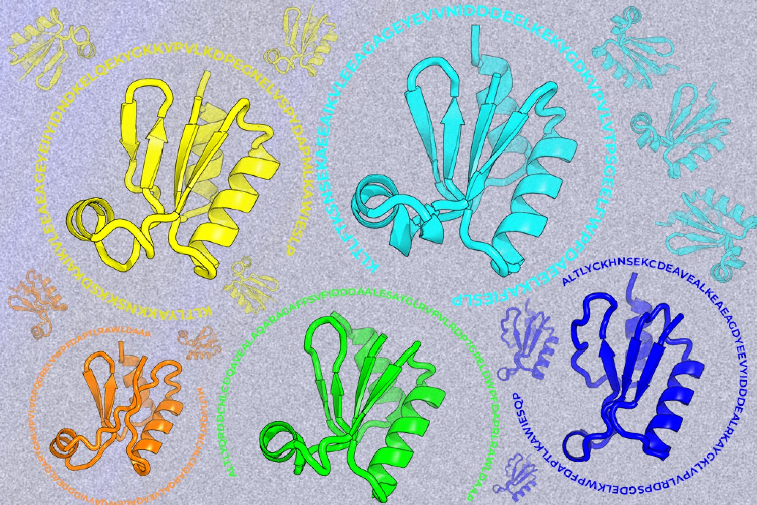
A new machine-learning framework aims to improve the success rate of computational protein design while moving away from results that reproduce sequences found in nature.