Aug 2026· Molecular Microbiology· 0 citations· 40 references
Medicine
TL;DR
The results suggest that roles of CetZ1 in cell shape or potentially the organisation and structure of the cell poles influence the polar assembly of the motility machinery.

Abstract
The archaeal tubulin-like cytoskeletal protein CetZ1 is required for rod-cell morphogenesis during the development of motility in Haloferax volcanii. This is expected to improve swimming speed and directionality. Here, we found that deletion of cetZ1 or expression of a GTPase-defective mutant caused the expected defects in cell shape, and also resulted in a substantial defect in the assembly of the motility machinery, including the archaellum base marker protein ArlD1, the chemotaxis sensory array adapter CheW1, and signal transducer CheY. Interestingly, moderate overexpression of cetZ1 (by 2-3 fold) reduced motility and the assembly and polar placement of the motility machinery without detectably affecting the rod shape of motile cells. We also investigated deletion of the conserved paralog cetZ2, which caused no detected defects in swimming or rod shape. However, overexpression of cetZ2 caused mild hyper-motility, whereas the cetZ2 GTPase-defective mutant reduced motility; these effects were dependent on the presence of cetZ1, suggesting that the altered cetZ2 might interfere with CetZ1's role in motility. Finally, we saw that a functional CetZ1-mTurquoise2 fusion strongly localized at the poles of mature motile cells, where it partially co-localized with the motility machinery markers. Overall, our results suggest that roles of CetZ1 in cell shape or potentially the organisation and structure of the cell poles influence the polar assembly of the motility machinery.
ABSTRACT Penetration of the peptidoglycan (PG) layer by the nascent flagellar rod is a critical step in basal body assembly and has long been attributed to the acetylglucosaminidase activity of the flagellar rod cap protein FlgJ. Previous work in Salmonella enterica suggested that occasional preexisting openings in the PG layer allow some flagella to assemble in the absence of the FlgJ enzymatic activity. More recently, studies in Bacillus subtilis demonstrated that membrane mobility of nascent flagellar structures enables rod penetration without dedicated PG hydrolysis. Here, we revisited the requirement for FlgJ acetylglucosaminidase activity in S. enterica by testing whether inhibition of class 3 flagellar gene expression by the anti-σ28 factor FlgM contributes to the flagellation defect of FlgJ catalytic mutants. Consistent with previous studies, loss of FlgJ acetylglucosaminidase activity did not abolish flagellar assembly but instead reduced its efficiency, resulting in a heterogeneous population in which many cells assembled functional basal bodies and flagella. Deletion of flgM significantly increased both the proportion of flagellated cells and the number of flagellar filaments per cell, indicating that reduced class three gene expression contributes substantially to the observed defect. These findings support a model in which FlgJ enzymatic activity enhances the efficiency of local PG remodeling but is not essential for rod penetration or basal body assembly. Our results demonstrate that acetylglucosaminidase-independent flagellar rod growth occurs in S. enterica, although less efficiently than in organisms that lack FlgJ-like PG hydrolases, highlighting the evolutionary diversity of mechanisms that accommodate flagellar assembly through the bacterial cell wall. IMPORTANCE Bacterial flagella allow pathogens, such as Salmonella, to navigate complex environments and invade host cells. During flagellar assembly, the basal body must traverse the peptidoglycan layer, a step-long thought to require the dedicated cell wall-degrading activity of FlgJ. Here, we show that Salmonella can assemble functional flagella, even when the acetylglucosaminidase activity of FlgJ is genetically inactivated. Although these mutants produce fewer flagella per cell, the assembly pathway remains active, and enhanced σ28-dependent gene expression partially restores flagellation. These findings reveal that early steps in flagellar assembly are more diverse than previously thought and that differences in cell wall structure between gram-positive and -negative bacteria likely determine whether a cell wall hydrolyzing activity is required for flagellum assembly. Bacterial flagella allow pathogens, such as Salmonella, to navigate complex environments and invade host cells. During flagellar assembly, the basal body must traverse the peptidoglycan layer, a step-long thought to require the dedicated cell wall-degrading activity of FlgJ. Here, we show that Salmonella can assemble functional flagella, even when the acetylglucosaminidase activity of FlgJ is genetically inactivated. Although these mutants produce fewer flagella per cell, the assembly pathway remains active, and enhanced σ28-dependent gene expression partially restores flagellation. These findings reveal that early steps in flagellar assembly are more diverse than previously thought and that differences in cell wall structure between gram-positive and -negative bacteria likely determine whether a cell wall hydrolyzing activity is required for flagellum assembly.
Yann H. U. Chevance, M. Kinoshita, F. Chevance et al.· Journal of Bacteriology· 0 citations
The cytoskeleton, comprising intracellular filamentous structures composed of polymerized proteins, is crucial for the survival of both eukaryotes and prokaryotes. Although bacterial cytoskeletal proteins have diverged, they generally do not drive cellular motility. Spiroplasma, a genus of wall-less helical bacteria, swims by propagating a helicity-switching point (kink) along its cell axis. Unlike typical walled bacteria, whose motility depends on widespread motility machineries such as flagella and pili, Spiroplasma swimming is powered by the coordinated dynamics of five isoforms of bacterial actin MreB (SMreB1–5), which are grouped into three phylogenetic classes: SMreB1 and 4, SMreB2 and 5, and SMreB3. Despite the efforts to understand Spiroplasma swimming, its molecular mechanism remains unclear. In this review, we summarize how in vitro analyses of SMreBs have provided mechanistic insights into Spiroplasma swimming. While all SMreBs conserve the canonical actin fold, each SMreB class exhibits unique characteristics in its polymerized structures, ATPase activities, polymerization dynamics, and membrane binding. Studies of an essential SMreB subset for Spiroplasma swimming, i.e. SMreB1 and SMreB5, have revealed that SMreB1 binds to polymerized SMreB5 and disassembles it depending on the nucleotide state. These results challenge the previous model in which Spiroplasma swimming is driven by the coordinated extension and contraction of two distinct SMreB filaments. Finally, we discuss potential molecular mechanisms underlying Spiroplasma swimming and highlight key questions that must be answered to validate these models.
Despite growing interest in the MreBCD morphogenetic complex as a potential antimicrobial target, its function in Pseudomonas aeruginosa remains poorly understood. While previous studies using the MreB inhibitor A22 have established its role in cell shape maintenance and pilus regulation, the impact of mreB deletion has not been comprehensively investigated. Using genetic and microscopy-based approaches, we show that deletion of mreB is viable in P. aeruginosa, but results in spherical cells that lose all forms of motility despite retaining flagella. Importantly, we uncover a previously overlooked polar effect of the in-frame mreB deletion on the downstream mreCD genes and show, using CRISPRi-mediated silencing, that mreCD expression is essential for viability. ΔmreB mutants also display increased sensitivity to β-lactam antibiotics and enhanced initial surface attachment, yet form more compact biofilms with reduced dispersal. In mixed-culture biofilms, spherical ΔmreB cells are outcompeted by rod-shaped wild-type cells and remain confined to the biofilm base. The identification of natural P. aeruginosa isolates carrying truncated mreB alleles further indicates that loss of MreB function can be tolerated in natural populations. Together, our findings reveal important contributions of the MreBCD system to viability, morphogenesis, motility and biofilm development in P. aeruginosa, providing new insights into bacterial adaptation and informing the development of targeted antimicrobial strategies.
M. Tunç, Mattéo Gérard, A. Barbotin et al.· bioRxiv· 0 citations
Eukaryotic cells change their shapes, actively segregate their DNA, and contain membrane networks, facilitated by a complex cytoskeleton containing actin filaments, microtubules made from tubulin, and other components. These filaments have ancient evolutionary origins because actin- and tubulin-like proteins form prokaryotic cytoskeletons in archaea and bacteria. Bona fide eukaryotic F-actin can be traced back to crenarchaea and Asgard archaea, which are the closest known relatives of eukaryotes. A possible Asgard archaeal origin of microtubules was suggested recently with the discovery of a lokiarchaeon containing AtubAB mini microtubules that share architectural features with their eukaryotic counterparts. Using phylogenetic analyses of metagenomic data, here we report the broad occurrence of tubulins in Asgard archaea. Biochemical and structural analyses showed that one of our previously unidentified heimdallarchaeial AtubAB tubulin pairs forms four-protofilament mini microtubules that show dynamic instability and are inhibited by the tubulin drug maytansine. Our work raises the possibility that microtubule architecture and dynamics evolved in Asgard archaea prior to eukaryogenesis.
Jan Löwe, Andriko von Kügelgen, V. J. Planelles-Herrero et al.· Science Advances· 0 citations
MreB, a bacterial actin homolog and polymerizing ATPase, is central to cell-shape maintenance and cell-wall integrity. Its functions rely on its ability to assemble into dynamic, membrane-associated polymers. However, how nucleotide binding and hydrolysis, MreB-MreB contacts, and membrane association are coordinated to enable polymer assembly and disassembly remains unclear. Here, we combined genetics and live-cell microscopy with biochemical approaches to dissect these processes. Using a highly sensitive reporter of MreB activity, we identified, through a genetic screen, residues critical for MreB function in Bacillus subtilis. Subsequent extensive characterization of corresponding stable variants of the homologous Geobacillus stearothermophilus MreB revealed that ATP binding, but not ATP hydrolysis, is required for polymerization. Productive longitudinal intraprotofilament contacts are required for efficient ATP hydrolysis and enhance membrane association. Perturbations predicted to weaken lateral interprotofilament contacts altered membrane association and modulated ATPase activity. Together, these effects provide experimental evidence consistent with long-range functional coupling among the longitudinal and lateral protofilament interfaces, the distant nucleotide-binding site, and membrane association dynamics. Moreover, impaired ATP hydrolysis delays disassembly of lipid-associated polymers, indicating that hydrolysis promotes polymer turnover. These results establish key mechanistic steps coordinating ATP-driven MreB polymerization and turnover and provide a basis for a complete MreB assembly–disassembly cycle and for further elucidating how MreB dynamics contribute to cell-wall organization.
Alba de San Eustaquio-Campillo, C. Cornilleau, Sana Afensiss et al.· bioRxiv· 0 citations
Related blog posts
MIT News · Artificial Intelligence· news.mit.eduAug 27, 2026
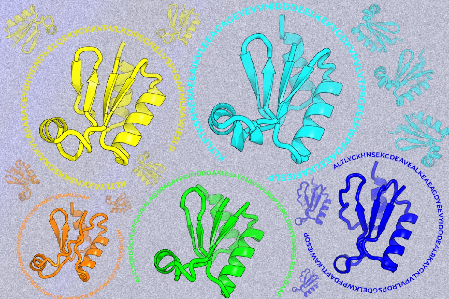
A new machine-learning framework aims to improve the success rate of computational protein design while moving away from results that reproduce sequences found in nature.