SLIM: Functional Nanoscopy for Simultaneous Super‐Resolution Imaging and Quantitative Lifetime Mapping by Spatiotemporal Multiplexing of Spontaneous Emission
Nov 2026· Laser & Photonics Reviews· 0 citations· 30 references
Abstract
Imaging nanoscale biological processes such as organelle interactions, protein aggregation, and metabolic regulation requires tools that concurrently resolve structural details and functional states. Stimulated emission depletion (STED)‐based super‐resolution can be integrated with fluorescence lifetime imaging (FLIM), but the depletion beam perturbs the fluorescence decay kinetics and compromises lifetime accuracy. Conventional FLIM, meanwhile, is fundamentally limited by optical diffraction. To overcome these limitations, we introduce SLIM, a functional nanoscopy technique based on spatiotemporal multiplexing of spontaneous emission. By combining a pulsed excitation laser with a temporally delayed STED depletion beam at an adjustable nanosecond interval, SLIM creates a temporal window within each acquisition cycle to separate spontaneous from stimulated emission, enabling the simultaneous acquisition of super‐resolved structural information and quantitative lifetime data. The technique delivers a 3‐ to 5‐fold improvement in structural resolution over diffraction‐limited FLIM, while preserving lifetime accuracy within 5% of confocal reference values, and operates at depletion powers one order of magnitude lower than conventional STED nanoscopy. We demonstrate SLIM's applicability across diverse biological specimens and its capability for long‐term live‐cell imaging of organelle dynamics. By merging structural imaging with quantitative functional mapping in a single modality, SLIM provides a practical and versatile platform for next‐generation biological discovery.
The combination of stimulated emission depletion (STED) microscopy with fluorescence lifetime imaging microscopy (FLIM) offers a powerful route to functional super-resolution imaging, but remains technically challenging for lifetime-based sensing because of the complex interplay between stimulated emission, photon budg...
Julia Rubio-Loscertales, Jose Requejo-Isidro, Cristina Flors· Methods and Applications in...· 0 citations
Direct monitoring the multifaceted intracellular microenvironment is fundamental for deciphering localized cellular behavior and function. However, performing nanoscale spatiotemporal sensing of the dynamic physiochemical environment within a live cell has remained challenging, mainly limited by the lack of robust, bio...
Yu-Xian Lu, Yi-Bo Yang, Ke Xu et al.· Nature Communications· 0 citations
By overcoming the diffraction limit of light, super-resolution fluorescence microscopy has become an indispensable tool for visualizing subcellular structures and dynamic biological processes. Nevertheless, conventional super-resolution techniques generally require extensive spatial or temporal sampling, resulting in p...
Förster resonance energy transfer (FRET) is a powerful tool for in vivo and in vitro studies of protein-protein interactions and conformational changes of complex biological molecules. For FRET studies in live cells and tissues, fluorescence lifetime imaging microscopy (FLIM) is advantageous due to the combined spatio-...
Gabriela Núñez-Gotia, Isaac Hanauska, Rylee Rosenberg et al.· Ultrafast Nonlinear Imaging...· 0 citations
Two‐photon microscopy (TPM) has revolutionized biological imaging by enabling high‐resolution, deep‐tissue visualization with reduced photobleaching and photodamage. This review discusses complementary nonlinear optical imaging modalities such as two‐photon fluorescence (TPF), second‐harmonic generation (SHG), and fluo...
Kausalya Neelavara Makkithaya, Gagan Raju, Subir Das et al.· Journal of Biophotonics· 0 citations
Related blog posts
MIT News · Artificial Intelligence· news.mit.eduNov 9, 2026
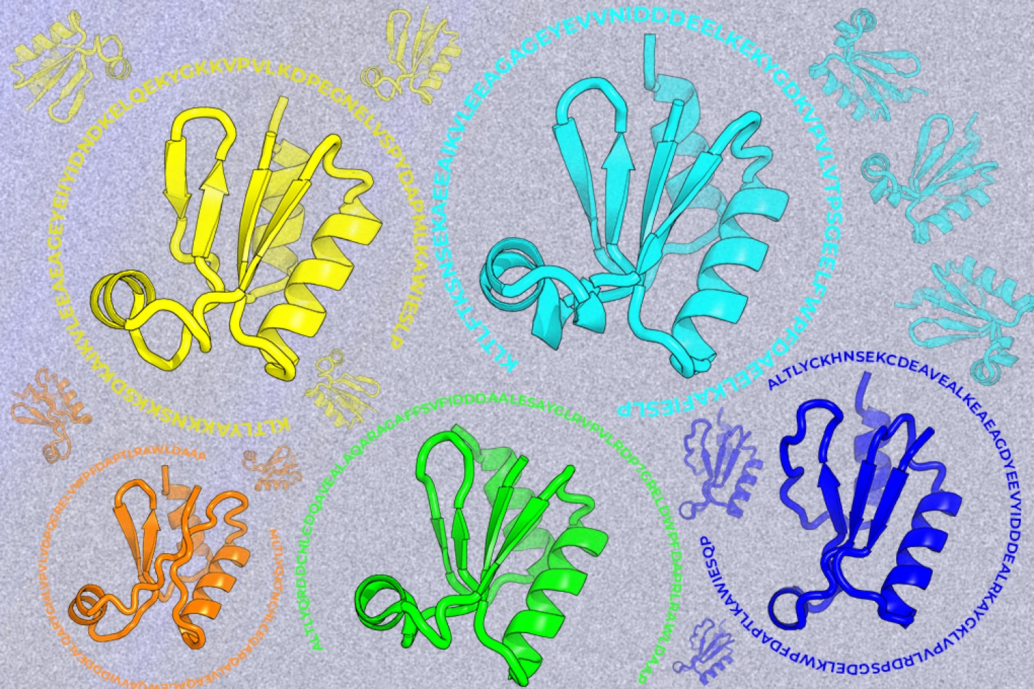
A new machine-learning framework aims to improve the success rate of computational protein design while moving away from results that reproduce sequences found in nature.