This study provides a practical framework for creating intrafilamentary porosity into 3D-printed PCL scaffolds with improved surface-mediated biological performance.

Abstract
Extrusion-based three-dimensional printing enables the fabrication of patient-specific scaffolds. Polycaprolactone (PCL)-based scaffolds have been widely used for the treatment of bone defects. However, extruded filaments are typically nonporous, which can prolong their degradation, limit cell adhesion and infiltration, and therefore interrupt normal tissue regeneration. Here, we present a simple and scalable method to generate polymeric scaffolds with hierarchical porosity by incorporating sacrificial sugar particles in a printable PCL ink. Microstructural analysis by SEM demonstrates an average pore size of around 10 μm with increased pore density at higher porogen fractions. Porous PCL improved apparent wettability and protein adsorption, accelerated hydrolytic degradation by more than five-fold, and enabled tunable mechanical properties. Printability testing shows improved filament stability and reduced fusion at higher porogen loading, enabling significantly higher printing resolution. In vitro evaluation demonstrates enhanced early preosteoblast attachment on porous scaffolds (>three-fold), without loss of cytocompatibility. Importantly, porous scaffolds support osteogenic differentiation of the cells and mineralization. This study provides a practical framework for creating intrafilamentary porosity into 3D-printed PCL scaffolds with improved surface-mediated biological performance.
Three-dimensional, interconnected hydrogel networks are central to tissue engineering and disease modeling, where tailored pore architecture and mechanical robustness are essential for supporting cellular functions. However, the limited ability to engineer microstructural features in vat polymerization 3D-printed natural hydrogels often compromises scaffold performance, as oversized pores reduce cell attachment and cell-cell interactions while smooth pore walls lack essential topographical cues. Here, we report an emulsion-based ink for vat polymerization 3D printing that enables the fabrication of hydrogels with finely tunable and highly interconnected porous architectures. An oil-in-water resin formulated using gelatin methacrylate (GelMA) contains stable solvent nanodroplets that act as sacrificial templates during photopolymerization. Removal of the dispersed phase yields additive-free porous hydrogels with pore sizes ranging from 0.66 to 46.15 µm and a 2.5-fold enhancement in compressive toughness. This strategy is compatible with digital light processing (DLP) and broadly applicable to multiple photocurable biopolymers, including alginate methacrylate (ALMA) and hyaluronic acid methacrylate (HAMA). The resulting porous scaffolds promote enhanced cell attachment, proliferation, and cell-cell interactions, highlighting the potential of this vat polymerization-compatible platform for advanced biofabrication.
Liwei Liu, Liwen Zhang, Xumin Huang et al.· Small Methods· 0 citations
Tissue engineering scaffolds (TESs) play a crucial role in regenerative medicine by providing structural support for cell adhesion, proliferation, differentiation, and tissue formation. However, developing TESs that simultaneously meet the requirements of biocompatibility, mechanical robustness, structural controllability, and cost-effective manufacturing remains a significant challenge. In this study, fully bio-based TESs were fabricated using polylactic acid (PLA) reinforced with cellulose nanofibers (CNFs) via a green and scalable microcellular injection molding process. The incorporation of CNFs derived from renewable biomass, significantly enhanced the rheological property, crystallinity, and foaming behavior of PLA. Compared with the PLA foams fabricated by regular foam injection molding (RFIM), the pore size of the PLA/CNF foam fabricated by mold-opening foam injection molding (MOFIM) was decreased by 96.5%, with the pore density increased by 7 orders of magnitude. The tensile toughness and impact strength were improved by up to 276.5% and 40.0%, reaching 6.4 MJ/m3 and 2.1 kJ/m2, respectively. Thanks to the improved scaffold architecture and introduced hydroxyl groups, the PLA/CNF foam enabled outstanding cell viability and proliferation, as evidenced by abundant live cells, uniform distribution, and minimal cell death. This work provides a sustainable and scalable strategy for developing high-performance TESs with tunable pore structures for biomedical applications.
Three dimensional (3D)-printed polylactic acid (PLA) scaffolds have gained significant attention for bone tissue regeneration due to their excellent biocompatibility, tunable architecture, and mechanical strength. However, the hydrophobic and bioinert surface of PLA limits its interaction with cells. To overcome these limitations, in this study, a multistep surface modification strategy is employed by combining 3D printing and electrospinning techniques. Here, 3D-printed PLA scaffolds are first treated with oxygen plasma to generate OPLA scaffolds with enhanced hydrophilicity, followed by coating with alginate (Alg)/poly(ethylene oxide) (PEO)-based electrospun nanofibers incorporated with magnesium phosphate nanoparticles (MP NPs) to develop Alg/PEO/MP/OPLA scaffold. Here, an extracellular matrix (ECM)-inspired electrospun nanofibrous coating provides a biomimetic surface for cell attachment, while poly(ethylene oxide) (PEO) improves the electrospinnability of alginate to obtain uniform nanofibers. Additionally, incorporation of MP NPs in nanofibers offers bioactive cues associated with osteogenic stimulation for bone regeneration. The successful coating of electrospun nanofibers on 3D-printed scaffolds is confirmed by attenuated total reflectance-Fourier transform infrared spectroscopy (ATR-FTIR) and field emission scanning electron microscopy (FE-SEM). Subsequent in situ mineralization results in calcium phosphate (CaP) deposition on the scaffold (Alg/MP/OPLA/CaP), as confirmed by ATR-FTIR, X-ray diffraction (XRD), and FE-SEM analysis. Further, in vitro cell studies demonstrate that the presence of MP NPs in Alg/MP/OPLA/CaP scaffold significantly improves cell attachment and proliferation over time, combined with enhancement in osteogenic activity. Overall, the developed Alg/MP/OPLA/CaP scaffold provides a bioactive environment for bone regeneration, thus serving as a potential alternative for the repair of critical-sized bone defects.
Critical-size bone defects (CSDs) remain a major clinical challenge. Although three-dimensional (3D) printing enables precise structural control and defect-specific scaffold fabrication, conventional polymer-based scaffolds often exhibit insufficient mechanical strength and limited osteogenic activity. In this study, poly(ε-caprolactone)/β-tricalcium phosphate (PCL/β-TCP) scaffolds containing 0, 1, 2, 4, or 8 wt% barium titanate (BaTiO3, BT) were fabricated by direct ink writing. Their rheology, microstructure, mechanical properties, local electromechanical response, cytocompatibility, angiogenesis-related activity, osteogenic differentiation, and bone regeneration in vivo were evaluated. All formulations exhibited suitable printability and regular porous structures. Moderate BT incorporation increased surface roughness and mechanical strength, with compressive strength remaining within the range of human cancellous bone. Piezoresponse force microscopy confirmed a detectable but mild local piezoelectric response. However, its direct osteogenic contribution was not isolated from other BT-related material effects and requires further verification. All scaffolds exhibited good cytocompatibility. PTB4 showed the most favorable biological performance, including enhanced MC3T3-E1 cell proliferation, increased ALP-positive area, greater mineralized deposition, and more pronounced VEGF-related fluorescence in HUVECs. After 12 weeks in a rat calvarial critical-size defect model, PTB4 exhibited significantly higher bone volume fraction and bone mineral density than the blank and PT groups. Histological and immunohistochemical analyses showed more extensive new bone formation, collagen deposition, and stronger osteogenesis-angiogenesis coupling. Overall, PTB4 achieved the best balance among printability, mechanical compatibility, cytocompatibility, angiogenesis-related activity, and osteogenic performance, supporting low-dose BT modification as a promising strategy for safe, printable scaffolds for critical-size craniofacial and oral bone defect repair.
Bone is a mineralized connective tissue composed of osteoblasts, osteocytes, and osteoclasts, and its integrity is essential for structural and physiological function. Defects arising from trauma, tumors, or developmental abnormalities often require surgical reconstruction to restore normal performance. Autografts and allografts have long served as standard treatments for bone repair, however, their usefulness is restricted by limited availability, donor‑site complications, and the potential transmission of underlying diseases. These challenges have accelerated interest in bone tissue engineering (BTE) as an alternative strategy capable of enhancing regeneration while reducing postoperative risks. Advances in three‑dimensional (3D) printing have introduced powerful technique for fabrication of scaffolds with precisely controlled architectures and tunable mechanical and biological characteristics. This technology enables the creation of porous constructs that mimic the structural complexity of native bone, supporting cell infiltration, nutrient transport, and vascularization. Effective scaffolds for BTE must demonstrate biocompatibility, biodegradability, appropriate strength and stiffness, and the ability to promote osteogenesis and angiogenesis. Among natural polymers, alginate (Alg) has become a prominent candidate due to its inherent biocompatibility, degradability, abundance, low cost, and non‑immunogenic nature. Its versatility makes it suitable for developing customized 3D‑printed scaffolds. Additionally, bioactive glasses (BGs) are widely incorporated into composite scaffolds because their composition closely resembles the mineral phase of bone. BGs significantly enhance osteoconductivity, support mineral deposition, and can improve the mechanical resilience of polymer-based constructs. This review highlights recent progress in 3D‑printed Alg-based scaffolds for BTE, emphasizing how advanced fabrication techniques and BGs incorporation contribute to improved biological performance and structural reinforcement.
Afsaneh Jahani, M. H. Ebrahimzadeh, Ali Moradi et al.· The Archives of Bone & Joint...· 1 citation
Related blog posts
MIT News · Artificial Intelligence· news.mit.eduAug 27, 2026
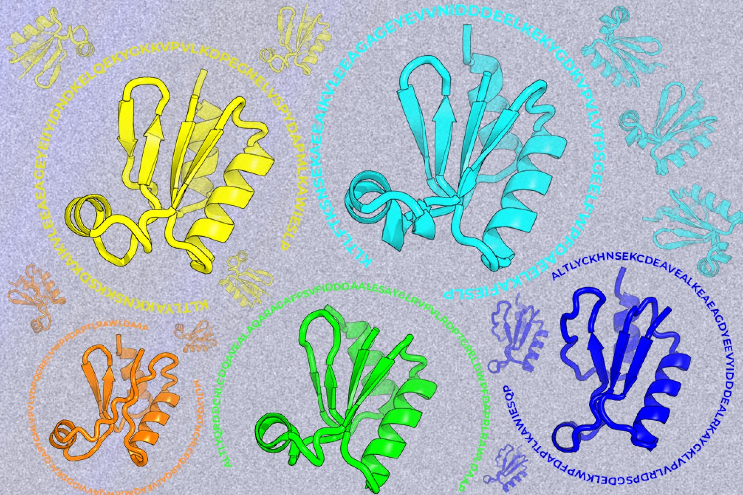
A new machine-learning framework aims to improve the success rate of computational protein design while moving away from results that reproduce sequences found in nature.